Posts

Bone marrow Biopsy

Image
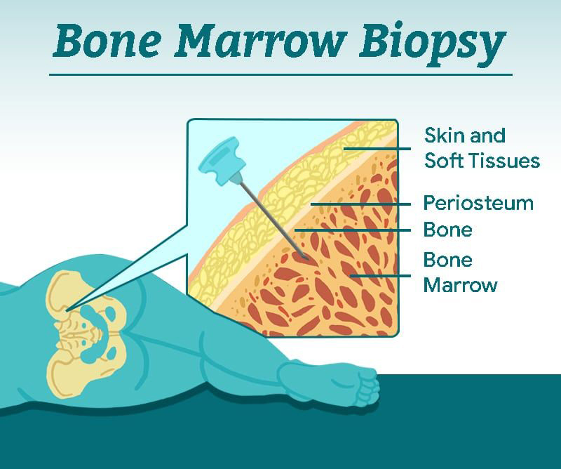
  Bone marrow biopsy (BMB) FAQs! Ever wondered what it's all about? Keep reading... 👀 What makes up a BMB? Usually it includes 2 samples - the aspirate (liquid marrow ðŸĐļ) and the trephine (bone core ðŸĶī) Why do we need 2 samples? The aspirate allows us to individually look at each BM cell under the microscope 🔎 (e.g. WBCs, RBCs) and classify them via flow cytometry (a method of identifying and measuring pathological cells using their surface markers e.g. CD19 for B-cells) The trephine lets us look at the way the BM is organised and assess for things like fibrosis and infiltration. We can also apply stains 🖌 (immunohistochemistry) to detect specific cells on the trephine How is it done? It is a quick (20-30 min) awake procedure using local anaesthetic. It does not require a theatre and is often done at the patient bedside. It is done aseptically using 2 separate needles 💉 (aspirate and trephine). Where do you take the samples from? Most times we manage to get samples from the pos...

BLOOD CANCER – TYPES AND TREATMENT

Image
Blood Cancer – Types and Treatment Blood accounts for about 8% of our total body weight and plays a vital role in bodily functions; circulating throughout body supplying nutrients, oxygen, antibodies to each and every cell.  Blood cancer occurs due to abnormal multiplying of leukocytes (WBCs). Cancer causes an imbalance in the body due to increase in cell division, leading to excessive production of WBCs. There are three main types of blood cancers: 1. Leukemia: Cancer affecting WBCs or precursor of white blood      cells, increasing their production that can’t fight infections. This      cancer generally originates in the bone marrow rendering its      ability of red blood cells and platelets production. It is further      categorized in four types: • Acute Lymphocytic Leukemia (ALL): As the name suggests, it     affects lymphocytes in the bone marrow. Production of too many     lymphocytes r...

REASONS FOR MUCORMYCOSIS IN COVID-19

Image
  Key Take Home Messages: §  Mucormycosis is an opportunistic devastating fungal rhino-cerebro-orbital infection which is primarily a disease of sinuses and secondary to orbit. §  Mucormycosis are not black fungi. It is not contagious & not spread by oxygenation, humidifier and water. §  Reasons for mucormycosis in Covid-19: v  affection of innate immunity v  oxidative stress v  iron overload v  affection of adaptive immunity §  Risk factors for mucormycosis in Covid-19: v  uncontrolled DM v  high dose and early treatment with steroids/immune-modulators/mechanical ventilation v  long standing oxygen therapy v  tocilizumab use v  prolonged use of broad spectrum antibiotics v  high levels of HbA1c, IL-6 and serum ferritin. §  Prevention: v  Good and proper hygiene of face and oral/buccal cavity v  No role of dehumidifier bottle fluid in this scenario, so no role of  methylene blue in these b...

MICROCYTIC AND HYPOCHROMIC ANEMIA IS ONE OF THE COMMON SCENARIO WE ENCOUNTER IN CLINICS

Image
  Clinical Pearls  #DrRajeshBollam  #Hematologist  Microcytic and Hypochromic anemia is one of the common scenario we encounter in Clinics  When we order iron studies  The following are possibilities and Probable diagnosis  *microcytic anaemia with low s.ferritin& high TIBC=iron defeciency anaemia *microcytic anaemia with high s.ferritin&low TIBC=anaemia of chronic diseases *microcytic anaemia with high s.iron= sidroblastic anaemia *microcytic anaemia with NORMAL iron studies=thalassemia  #Hematologist #Hyderabad #Telangana #DrRajeshBollam #anemia BOOK APPOINTMENT

UPDATED RECOMMENDATIONS FOR BREAST CANCER SURVEILLANCE IN YOUNG FEMALE CANCER SURVIVORS

Image
  Updated Recommendations for Breast Cancer Surveillance in Young Female Cancer Survivors William J. Gradishar, MD reviewing Mulder RL et al. J Clin Oncol 2020 Dec 10 Surveillance is now strongly recommended for survivors treated with ≥10 Gy chest radiation — a change from ≥20 Gy. In 2013 the International Late Effects of Childhood Cancer Guideline Harmonization Group (IGHG) published evidence-based recommendations for surveillance of female survivors of childhood, adolescent, and young adult (CAYA) cancer who were treated with radiation to fields that potentially involved breast tissue on the chest wall. Based on new evidence, IGHG has updated its breast cancer surveillance recommendations. BOOK APPOINTMENT Modified recommendations include the following: Mammography and breast MRI should be performed at least annually up to age 60 for survivors treated with ≥10 Gy chest radiation (strong recommendation) and for those treated with upper abdominal radiation that exposed breast tiss...

Risk Factors of Lung Cancer

Image
Risk factors for lung cancer The biggest risk factor for lung cancer is smoking. That includes cigarettes, cigars, and pipes. Tobacco products contain thousands of toxic substances. According to the Centers for Disease Control and Prevention (CDC) Trusted Source, cigarette smokers are 15 to 30 times more likely to get lung cancer than nonsmokers. The longer you smoke, the greater the chance of developing lung cancer. Quitting smoking can lower that risk. Breathing in secondhand smoke is also a major risk factor. Every year in the United States, about 7,300 people who’ve never smoked die from lung cancer caused by secondhand smoke. Exposure to radon, a naturally occurring gas, increases your risk of lung cancer. Radon rises from the ground, entering buildings through small cracks. It’s the leading cause of lung cancer in nonsmokers. A simple home test can tell you if the level of radon in your home is hazardous. Your risk of developing lung cancer is higher if yo...

Diagnosing lung cancer

Image
After a physical examination, your doctor will tell you how to prepare for specific tests, such as: Imaging tests : An abnormal mass can be seen on X-ray, MRI, CT, and PET scans. These scans produce more detail and find smaller lesions. Sputum cytology : If you produce phlegm when you cough, microscopic examination can determine if cancer cells are present. A biopsy can determine if tumor cells are cancerous. A tissue sample can be obtained by: Bronchoscopy : While under sedation, a lighted tube is passed down your throat and into your lungs, allowing closer examination. Mediastinoscopy : The doctor makes an incision at the base of the neck. A lighted instrument is inserted and surgical tools are used to take samples from lymph nodes. It’s usually performed in a hospital under general anesthesia. Needle : Using imaging tests as a guide, a needle is inserted through the chest wall and into th...